New MRI Technology Shows the Brain 64 Million Times Clearer
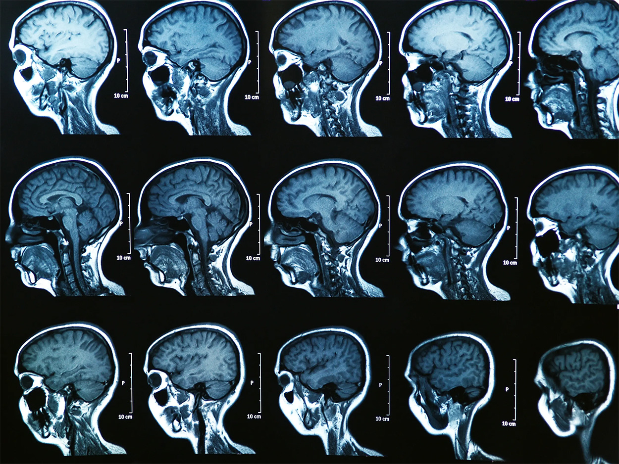
Fifty years ago, MRI or magnetic resonance imaging debuted in the market, developed by Pal Laterbur, an American chemist. On its 50th anniversary, scientists from Duke University and other collaborators marked the historical day with the announcement of the sharpest scans of a brain. However, it is the scan of a lab mouse’s brain, at least for now.
Years of work
It took researchers from the Center In Vivo Microscopy of Duke University, together with scientists from Indiana University, the University of Pittsburgh, the University of Pennsylvania, and the University of Tennessee Health Science Center close to 40 years to perfect the MRI that can generate scans that are 64 million times sharper than current MRI machines.
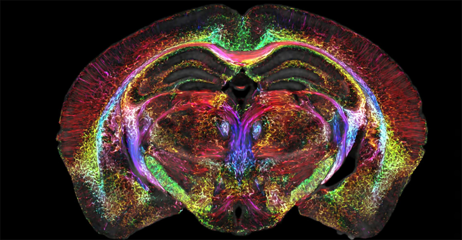
The technology, which the researchers call HiDiver, short for high-dimensional integrated volume with registration, captures highly detailed images. Each voxel, or a pixel’s 3D version, is only about five microns. Although the current MRI technology is already advanced to find a brain tumor, the HiDiver technology can display organization and more detailed connectivity and circuitry data to help doctors identify issues in the brain.
Excellent detailed imaging
The research team believes the detailed imaging HiDiver achieves will better understand how brains change with diet, neurodegenerative diseases, and age.
Lead author G. Allan Johnson, a Duke University professor of radiology, physics, and biomedical engineering, calls their work enabling because it allows them to look at neurodegenerative diseases like Alzheimer’s in an entirely different way.
Use of impressively advanced technology
The researchers worked at the Center for In Vivo Microscopy for nearly 40 years, using the most impressive technology to create the MRI resolution they envisioned. Clinical MRIs today have 1.5 to 3-Tesla magnets. They equipped the HiDiver with a 9.4-Tesla magnet, a set of gradient coils 100 times stronger than the standard scans, and a supercomputer with the power of 800 laptops. These components unite to capture the single mouse brain in their test.
After completing the MRI visual, they scanned the mouse brain tissue using light sheet microscopy, enabling them to label particular cell groups, which let them watch the progress of neurodegenerative diseases over time.
They made their study more conclusive by using different sets of mice in different genetic makeups and ages, allowing them to see how brain-wide connectivity changes over time. It also showed them how specific regions, such as the memory-related subiculum, can change more than other brain parts. HiDiver likewise captured images showing how neural networks break down due to Alzheimer’s.
According to the team, their work opens the path for further technological development for capturing images of the human brain in greater detail. The technology will provide researchers and doctors further understanding of how brain tissue change with age. It would also help them to know what interventions they can provide to avert degeneration.
Rising to the challenge
The research collaborators believe that their new brain imaging technique, which can produce images 64 million times sharper than the MRI machines used today, can change the medical field’s understanding of how neurodegenerative diseases like Alzheimer’s, Parkinson’s disease, Friedreich ataxia, amyotrophic lateral sclerosis, Huntington’s disease, and spinal muscular atrophy, progress.
They took on the challenge to study one of the body’s most complex organs, the brain. The human brain is about the size of two fists, but it contains 86 billion neurons, which form 100 trillion connections with one another. Thus, it is extremely difficult to understand what leads to brain disorders and develop effective treatments for these neurodegenerative diseases.
With the HiDiver MRI technology, researchers can now look at these brain diseases, and with the results of their study of the mouse brain, they can soon use the technology in humans.